Cycle Science: Ovulation, Luteal Phase & Due Dates
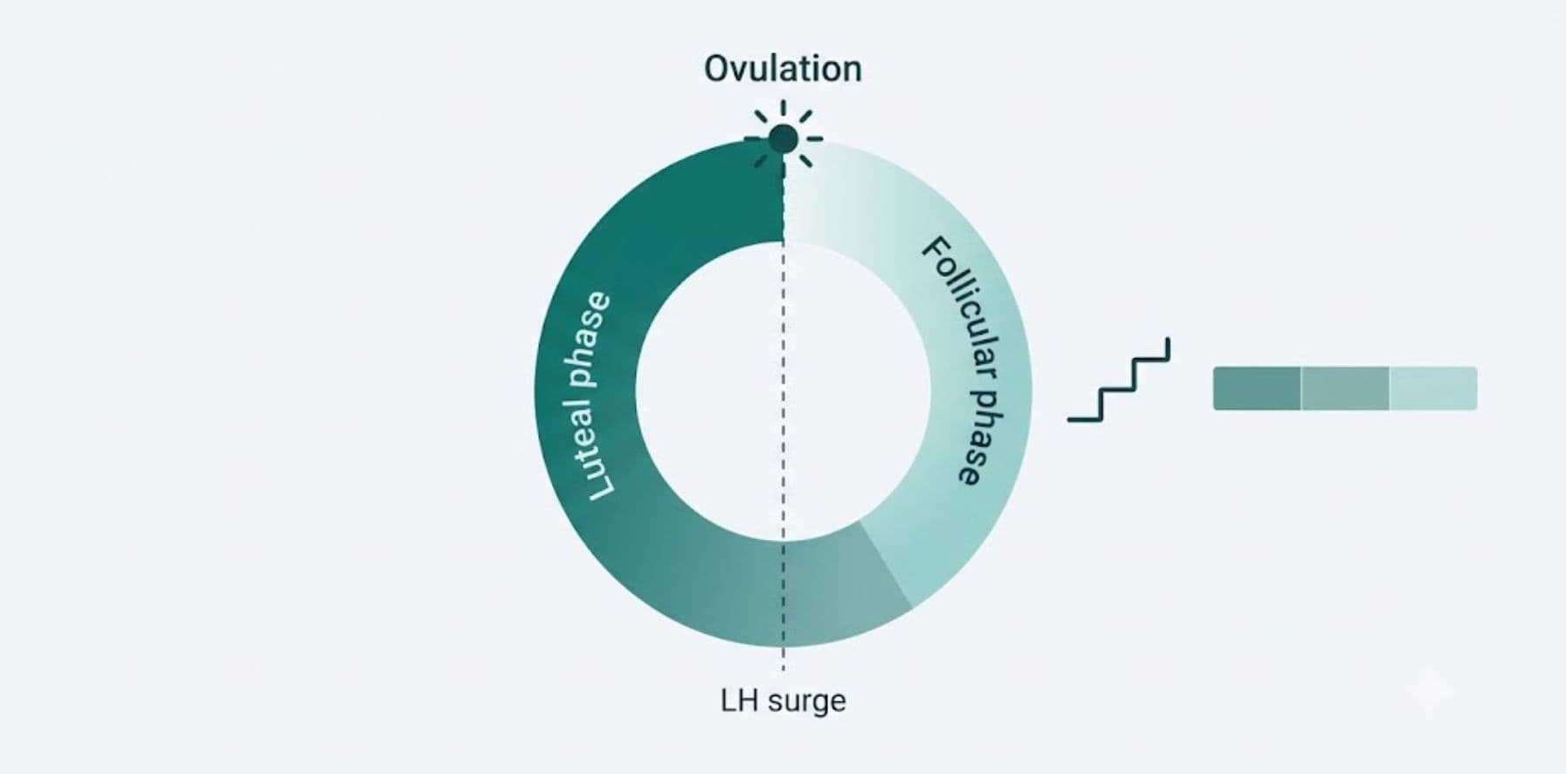
Menstrual cycle tracking has evolved far beyond a paper calendar with circled dates. Modern predictive algorithms model the interplay of the hypothalamic-pituitary-ovarian (HPO) axis, hormone half-lives, and individual cycle variance to project ovulation windows and gestational timelines with clinical precision. This guide breaks down the endocrinology behind those projections the follicular dynamics, gonadotropin surges, and luteal phase consistency that any accurate Reproductive Health Tracking Suite must account for.
Foundations of Menstrual Physiology: The Three-Phase Model
A menstrual cycle is not a single uniform event it is a sequence of three distinct, hormonally governed phases: the follicular phase, ovulation, and the luteal phase. Cycle length is conventionally measured from Day 1 (first day of menstrual bleeding) to the day before the next period begins. While the textbook average is 28 days, clinically normal cycles range from 21 to 35 days, and length variability of ±2–3 days between cycles is considered physiologically unremarkable.
Follicular Phase Dynamics
The follicular phase spans from Day 1 until ovulation and is the most variable segment of the cycle in length. It begins with a drop in estrogen and progesterone, which triggers the hypothalamus to release gonadotropin-releasing hormone (GnRH) in pulsatile bursts. This stimulates the anterior pituitary to secrete follicle-stimulating hormone (FSH), recruiting a cohort of ovarian follicles. Through a process called follicular selection, one dominant follicle typically outcompetes the others by Day 7–10, continuing to mature while producing rising levels of estradiol. This estrogen rise is what triggers the endometrial proliferative phase thickening the uterine lining in preparation for potential implantation.
Diagnostic Snapshot: Follicular Phase Markers
- Duration: 10–22 days (most variable phase; shortens with age)
- Dominant hormone: Estradiol (rising trajectory)
- Pituitary signal: FSH pulse frequency ~1 pulse/1–2 hours early phase
- Endometrial state: Proliferative glandular re-epithelialization
- Basal body temperature: Low, stable baseline (~97.0–97.5°F / 36.1–36.4°C)
Ovulatory Phase & the Gonadotropin Spike
Once estradiol from the dominant follicle crosses a sustained threshold (typically >200 pg/mL for approximately 48 hours), it flips from suppressing to stimulating the pituitary a positive feedback loop that triggers the luteinizing hormone (LH) surge. This gonadotropin spike is the single most reliable biochemical predictor of ovulation: the LH surge precedes follicular rupture by approximately 24–36 hours, making it the biomarker of choice for urine-based ovulation predictor kits (OPKs). Ovulation itself the release of the mature oocyte from the follicle occurs roughly 10–12 hours after the LH peak.
Many individuals also experience Mittelschmerz, a German term meaning "middle pain," referring to unilateral lower abdominal discomfort correlating with follicular rupture. Clinically, the Mittelschmerz symptom array typically includes:
- Sharp or cramping unilateral pelvic pain, lasting minutes to 48 hours
- Mild spotting or increased cervical discharge (egg-white, high spinnbarkeit consistency)
- A measurable basal body temperature (BBT) shift of +0.4–1.0°F (0.2–0.5°C) post-ovulation, caused by progesterone's thermogenic effect
- Transient bloating or breast tenderness as progesterone begins to rise
Ovulation Biomarker Hierarchy (Reliability, High → Low)
- Serum LH / progesterone lab draw (gold standard, retrospective confirmation)
- Urinary LH surge (OPK) 24–36 hour predictive window
- BBT thermal shift confirms ovulation retrospectively, not predictive
- Cervical mucus consistency (Billings/ovulation method)
- Mittelschmerz and secondary somatic symptoms supportive, non-diagnostic
For a composite reading that cross-references cycle-day probability against biomarker input, the Ovulation & Fertility Calculator layers these signals rather than relying on calendar math alone.
Luteal Phase Consistency: The Predictive Anchor
Unlike the follicular phase, the luteal phase from ovulation to the onset of the next menses is remarkably consistent within an individual, which is precisely why it functions as the stable anchor point in cycle-prediction algorithms. The ruptured follicle transforms into the corpus luteum, a transient endocrine structure secreting progesterone (and some estradiol) to sustain the secretory endometrium. Absent fertilization and subsequent human chorionic gonadotropin (hCG) rescue signaling, the corpus luteum regresses at approximately 12–14 days, progesterone withdraws, and menstruation begins.
Clinical literature places the luteal phase at 12–14 days with a standard deviation of roughly ±1–2 days dramatically tighter than the follicular phase's variance. This asymmetry is why accurate cycle-prediction algorithms work backward from the expected next period using a fixed luteal length, rather than simply projecting forward from Day 1, and it is the core mathematical principle underlying the Period Tracker & Predictor.
Predictive Modeling: The Mathematics of Cycle Forecasting
Fertility-window windowing the practice of estimating a probable conception interval combines two data points: average cycle length and luteal phase stability. Because sperm can survive in the reproductive tract for up to 5 days and the oocyte remains viable for roughly 12–24 hours post-ovulation, the fertile window is modeled as a 6-day span: the 5 days preceding ovulation plus the day of ovulation itself, with peak conception probability concentrated in the 2 days immediately before ovulation.
Core Formula: Estimated Ovulation Day (EOD)
EOD = Next Predicted Period Start − Average Luteal Phase Length (typically 14 days)
Example: A 30-day average cycle with a 14-day luteal phase places ovulation at approximately Day 16 not Day 15, which is the common miscalculation produced by naive midpoint (cycle length ÷ 2) estimation. The midpoint method only holds true for a textbook 28-day cycle; any deviation requires luteal-phase-anchored back-calculation for accuracy.
Naegele's Rule: The Foundational Due Date Formula
Once conception is confirmed, gestational age estimation shifts from ovulation-tracking to due-date projection. The most widely used clinical formula remains Naegele's Rule, developed in the 19th century and still the baseline reference in obstetric practice for calculating an estimated due date (EDD):
Naegele's Rule
EDD = LMP (Last Menstrual Period start date) + 7 days − 3 months + 1 year
This formula assumes a standard 28-day cycle with ovulation on Day 14 and a 280-day (40-week) gestation measured from LMP not from the actual conception date, which is typically 2 weeks later. This is why obstetric "gestational age" and true "embryological/fertilization age" differ by roughly 14 days.
For cycles that deviate from 28 days, clinicians apply a correction: add (average cycle length − 28) days to the Naegele output. A 32-day average cycle, for instance, shifts the EDD 4 days later than the unadjusted formula would suggest a nuance the Pregnancy Due Date & Milestones tool factors in automatically rather than defaulting to the textbook assumption.
Ultrasound Dating vs. LMP Dating
Where first-trimester ultrasound (crown-rump length, CRL) is available, ACOG guidance favors sonographic dating over LMP-based Naegele calculation whenever the discrepancy exceeds 5–7 days in the first trimester, since CRL measurement carries a margin of error of only ±3–5 days versus LMP recall error, irregular cycle length, or anovulatory bleeding that can meaningfully skew Naegele's output.
Gestational Age Estimation & Trimester Progression
Gestational age is conventionally expressed in completed weeks and days (e.g., "18 weeks 3 days" or "18w3d") counted from LMP. The 40-week gestation is divided into three trimesters, each with distinct physiological milestones relevant to both maternal monitoring and fetal development benchmarking.
Trimester Progression Timeline
First Trimester
Weeks 0–13 (0–13w6d)
Organogenesis; hCG doubling every 48–72 hours in early weeks; highest miscarriage-risk window; dating scan performed weeks 8–13.
Second Trimester
Weeks 14–27 (14–27w6d)
Anatomy scan at 18–22 weeks; quickening (first fetal movement) typically felt weeks 16–22; gestational diabetes screening weeks 24–28.
Third Trimester
Weeks 28–40+
Group B Strep screening weeks 36–37; term defined as 37w0d–41w6d; post-term risk monitoring begins 41 weeks.
Frequently Asked Questions
Why does the follicular phase vary more than the luteal phase? ▾
The follicular phase length depends on how quickly a dominant follicle is recruited and matures, which is sensitive to stress, illness, and age-related ovarian reserve decline. The luteal phase, by contrast, is governed by the fixed lifespan of the corpus luteum (~12–14 days), which degrades on a relatively constant hormonal timetable regardless of external factors making it the more reliable variable for backward-calculating ovulation date.
How accurate is Naegele's Rule compared to ultrasound dating? ▾
Naegele's Rule assumes a textbook 28-day cycle with Day-14 ovulation, so its accuracy declines for anyone with irregular or non-average cycle lengths. First-trimester CRL ultrasound has a margin of error of roughly ±3–5 days and is considered more reliable, which is why clinical guidelines favor ultrasound dating when it differs meaningfully from LMP-based calculation.
What's the difference between gestational age and fertilization age? ▾
Gestational age is counted from the first day of the last menstrual period, roughly two weeks before ovulation and conception actually occur. Fertilization (embryological) age counts from the actual conception date. This 14-day offset explains why a pregnancy is described as "40 weeks" from LMP despite the embryo only developing for about 38 weeks.
Can basal body temperature alone confirm ovulation before it happens? ▾
No. BBT shifts occur after ovulation, driven by progesterone's thermogenic effect from the corpus luteum, so a sustained temperature rise confirms that ovulation has already occurred rather than predicting it in advance. For predictive timing, the LH surge (via OPK) is the more clinically useful leading biomarker.
This content is provided for educational purposes and does not substitute for personalized medical advice. Individuals with irregular cycles, suspected PCOS, or fertility concerns should consult a licensed reproductive endocrinologist or OB-GYN.